

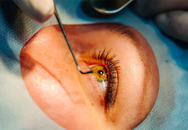
Retinal detachment is a serious eye condition in which the retina separates from the tissue supporting it at the back of the eye. If left untreated, it can cause permanent vision loss. Sudden flashes of light, new or increasing floaters, blurred vision, or a dark curtain-like shadow across your vision can be warning signs that need prompt medical attention.
Retinal detachment surgery is used to reattach the retina and protect vision when surgery is required. Depending on the type and severity of the condition, treatment may include pneumatic retinopexy, scleral buckling, vitrectomy, laser treatment, or cryotherapy.
This guide explains the symptoms, causes, diagnosis, treatment options, recovery, risks, and when to consult an ophthalmologist.
What Is Retinal Detachment?
The retina is the light-sensitive layer at the back of the eye. It converts light into signals that travel to the brain through the optic nerve, allowing us to see.
Retinal detachment occurs when the retina separates from the underlying tissue that provides it with oxygen and nutrients. The affected part of the retina may then stop functioning normally.
Because prolonged detachment can damage retinal cells, prompt assessment and appropriate retinal detachment treatment are important for protecting vision.
What Are the Early Signs of Retinal Detachment?
The early signs of retinal detachment may develop suddenly. Common warning signs include:
- Flashes of light in the eye
- A sudden increase in floaters
- Blurred or reduced vision
- Loss of peripheral vision
- A dark curtain or shadow across part of the visual field
These symptoms can also occur with other eye conditions. However, sudden flashes or numerous new floaters may indicate a retinal tear, which can sometimes progress to detachment.
If you experience sudden vision changes or a curtain-like shadow, seek prompt evaluation from an eye specialist.
What Causes Retinal Detachment?
A common type develops when a retinal tear or hole allows fluid to pass underneath the retina. This separates the retina from the tissue beneath it.
Factors that may increase the risk include:
- High myopia or severe short-sightedness
- Previous eye surgery
- Eye injury or trauma
- Previous retinal tear or detachment
- Certain retinal conditions
- Increasing age
Having a risk factor does not mean that retinal detachment will definitely occur.
How Is Retinal Detachment Diagnosed?
An ophthalmologist usually performs a detailed eye examination, often after dilating the pupils. This allows the retina to be examined for tears, holes, bleeding, and areas of detachment.
An eye ultrasound may be recommended when the retina cannot be clearly visualised or when additional assessment is required. The examination findings help determine the most appropriate treatment.
Does Retinal Detachment Always Need Surgery?
Many established retinal detachments require prompt treatment, and several cases require a procedure to reattach the retina. However, there is no single retinal detachment surgery that is suitable for every patient.
The choice depends on the location and extent of the detachment, the number and position of retinal tears, whether the macula is involved, and the overall condition of the eye.
An ophthalmologist will assess these factors before recommending a treatment approach.
What Are the Types of Retinal Detachment Surgery?
Scleral Buckling
Scleral buckling involves placing a silicone band or buckle around the outside of the eye. It supports the eye wall and reduces the forces that pull on the retina. Laser treatment or cryotherapy may also be used to seal retinal tears.
Vitrectomy
Vitrectomy for retinal detachment involves removing the vitreous gel from inside the eye. The surgeon can then treat retinal tears and reattach the retina.
A gas bubble or silicone oil may be used to provide internal support while the retina heals.
Pneumatic Retinopexy
Pneumatic retinopexy involves injecting a gas bubble into the eye. The bubble helps push the detached retina back into position, while laser or cryotherapy can seal the retinal tear.
This treatment is appropriate only for selected types and locations of retinal detachment.
What Is Recovery Like After Retinal Detachment Surgery?
Retinal detachment surgery recovery varies depending on the procedure, extent of the detachment, and individual healing.
Temporary blurred vision, redness, swelling, or mild discomfort may occur during the early recovery period. Eye drops may be prescribed, and patients should follow their ophthalmologist’s instructions about activity, eye protection, and follow-up appointments.
If a gas bubble is placed inside the eye, a specific head position may be recommended to support retinal healing.
Air travel may be unsafe while certain intraocular gas bubbles remain in the eye. Patients should not fly or travel to high altitudes until their eye surgeon confirms that it is safe.
Vision may improve gradually over several weeks or months. The final outcome can depend significantly on whether the macula was affected.
Can Vision Return After Retinal Detachment Surgery?
Vision recovery after retinal detachment surgery varies from patient to patient.
Successful reattachment does not always mean that vision immediately returns to its previous level. Factors such as macular involvement, the extent and duration of the detachment, and the overall health of the eye can influence visual recovery.
Your ophthalmologist can provide a more individualised prognosis after examining your eye.
What Are the Risks of Retinal Detachment Surgery?
As with other eye procedures, retinal surgery can have potential complications. Depending on the procedure and individual circumstances, these may include:
- Cataract development or progression
- Increased eye pressure or glaucoma
- Infection
- Bleeding
- Recurrent retinal detachment
- Persistent visual changes
- Need for additional treatment or surgery
Cataract and glaucoma are separate eye conditions that may also require evaluation and treatment. You can learn more about cataract and glaucoma through our detailed guides.
Following post-operative instructions and attending scheduled examinations are important for monitoring recovery.
When Should You See an Eye Specialist?
Do not ignore sudden flashes of light, a rapid increase in floaters, a dark curtain over your vision, or sudden loss of sight.
These symptoms may indicate a retinal tear or detached retina and require prompt assessment by an ophthalmologist. Dr. Y.R. Valentina, MBBS., MS(O), from the Department of Ophthalmology at Ponjesly Super Speciality Hospital, recommends seeking timely medical evaluation when such sudden vision changes occur. Early evaluation can help identify the condition and determine whether urgent treatment is needed.
Retinal Detachment Treatment in Nagercoil
If you are looking for retinal detachment treatment in Nagercoil, timely evaluation by an ophthalmologist is important when you experience sudden changes in vision.
The Ophthalmology Department at Ponjesly Super Speciality Hospital, Nagercoil provides evaluation and management for a range of eye conditions, including retina and vitreous diseases.
Patients are assessed based on their symptoms and clinical findings, and the appropriate treatment approach is recommended according to their individual condition.
If you are looking for the best eye hospital in Nagercoil for ophthalmology evaluation, contact Ponjesly Super Speciality Hospital to book a consultation.
Frequently Asked Questions
Is retinal detachment an emergency?
Yes. Suspected retinal detachment requires prompt ophthalmic assessment because delayed treatment can increase the risk of permanent vision loss.
What are the first signs of a detached retina?
Common warning signs include sudden flashes of light, new or increasing floaters, blurred vision, loss of peripheral vision, or a curtain-like shadow across the field of vision.
Does retinal detachment always require surgery?
Many retinal detachments require a procedure to reattach the retina, but the treatment depends on the type, location, and extent of the detachment and the findings during examination.
How long does retinal detachment surgery recovery take?
Recovery varies between patients. Vision may continue to improve for several weeks or months depending on the severity of the condition, treatment performed, and whether the macula was affected.
Can I fly after retinal detachment surgery?
If a gas bubble has been placed inside the eye, flying can be unsafe until the bubble has cleared. Always obtain clearance from your treating eye surgeon before air travel.
Can retinal detachment cause permanent vision loss?
Yes. Delayed treatment can result in permanent vision loss. Prompt evaluation and appropriate treatment are important when warning symptoms occur.
Need an Eye Examination?
Sudden flashes, new floaters or changes in vision should not be ignored. Get timely evaluation from an ophthalmologist.
Book an Appointment